Picture Of Animal Cell Under Microscope / a. Cell : unit of function - BIOLOGY4ISC / Wide collections of all kinds of labels pictures online.. Human animal cell under microscope. Huge collection, amazing choice, 100+ million high quality, affordable rf and rm images. Published on december 9, 2013 at 8:13pm by glenda stovall under cell. Place the glass slide onto the stage. Published by suhendra gunawan modified over 2 years ago.
This illustration is part of animal cell. The cells may be removed directly or by mechanical or enzymatic action. Even more amazing is to see your own cells under the microscope. Most cells, both animal and plant, range in size between 1 and 100 micrometers and are thus visible only with the aid of a microscope. They reveal complex patterns in creatures such as mice and fruit flies that are also.
Animal Cells Microscope Slide : Biological Science Picture ... from pulpbits.net Be careful pushing it under the clips that the cover slide doesn't move or crack. This illustration is part of animal cell. It is the outermost membrane of an animal cell having a thickness. Make your work easier by using a label. Cautionary labels are given for products or containers containing hazardous material. Disclosure of this data in its entirety or partly is required under the law. I am studying a bachelor's degree in biology, and i take a lot of pictures of what we do in the labs so i can remember what was everything like when i study. For example, the low power.
Cautionary labels are given for products or containers containing hazardous material.
Feel free to post your own, but please read the rules first (see below), and note that we are not a (1a) no screenshots or pics where the only focus is a screen. Wide collections of all kinds of labels pictures online. For example, the low power. Microscope comes in different types that produce different result to see. Animal cells • there are a number of differences between plant and animal cells when they are viewed under a microscope • cell size and shape of animal and plant cells differ • some organelles are found only in one cell type, but not in both (cell wall and chloroplast in plant cells. However, the internal structure and organelles are more or less similar. (reproduced by permission of photo. They reveal complex patterns in creatures such as mice and fruit flies that are also. Select the lowest power objective lens. Meiosis animal cell under microscope education stock photo edit. This picture of an animal cell was taken using an electron microscope. Published on december 9, 2013 at 8:13pm by glenda stovall under cell. Cells under microscope foto sin derechos de autor a generalised animal cell as observed under an electron microscope.
The cells may be removed directly or by mechanical or enzymatic action. This illustration is part of animal cell. A cell is a very tiny structure which exists in living bodies. Under the microscope, animal cells appear different based on the type of the cell. This picture of an animal cell was taken using an electron microscope.
The Cell - Form 1 Biology Notes from www.easyelimu.com With the correct staining or an in vivo tagging strategy, most organelles can be seen using light microscopy. Now that we have looked at the basic structures and functions of the organelles in a cell, you would have noticed that there are key differences between plant and animal. However, the internal structure and organelles are more or less similar. They reveal complex patterns in creatures such as mice and fruit flies that are also. However, it is best if the staining is specific to the particular organelle. Huge collection, amazing choice, 100+ million high quality, affordable rf and rm images. In this video, you will explore 3 different microscopic views of human. 4) cell membrane or plasma membrane.
The cells may be removed directly or by mechanical or enzymatic action.
Select the lowest power objective lens. Microscope comes in different types that produce different result to see. (reproduced by permission of photo. Published by suhendra gunawan modified over 2 years ago. The animal cell is more fluid or elastic or malleable in structure; They reveal complex patterns in creatures such as mice and fruit flies that are also. Vector illustration on a light background cells under a microscope. As being single layers, such cells can be transferred directly to a cover slip to examine under a microscope. Cautionary labels are given for products or containers containing hazardous material. Sponsored link you might be interest with others pictures of 6 animal cell microscope. Examining animal cells under the microscope. 7 in order to see all the organelles in a cell, you need a more powerful microscope. Under the microscope, animal cells appear different based on the type of the cell.
Cautionary labels are given for products or containers containing hazardous material. Vector illustration on a light background cells under a microscope. Each of these epithelial cells was examined under the microscope as students. Stunning images of life's building blocks under the microscope set to light up times square. Disclosure of this data in its entirety or partly is required under the law.
Labelled Diagram Of A Plant Cell Under A Microscope ... from www.smartsciencepro.com The images taken by lab scientists have helped biologists uncover new treatments for a range of diseases. Make your work easier by using a label. Removing cellular waste products from the cell. Most cells, both animal and plant, range in size between 1 and 100 micrometers and are thus visible only with the aid of a microscope. Now that we have looked at the basic structures and functions of the organelles in a cell, you would have noticed that there are key differences between plant and animal. Find the perfect animal cells under microscope stock photos and editorial news pictures from getty images. 7 in order to see all the organelles in a cell, you need a more powerful microscope. Generalized cell is used for structure of animal cell and plant cell to present the common parts.
As being single layers, such cells can be transferred directly to a cover slip to examine under a microscope.
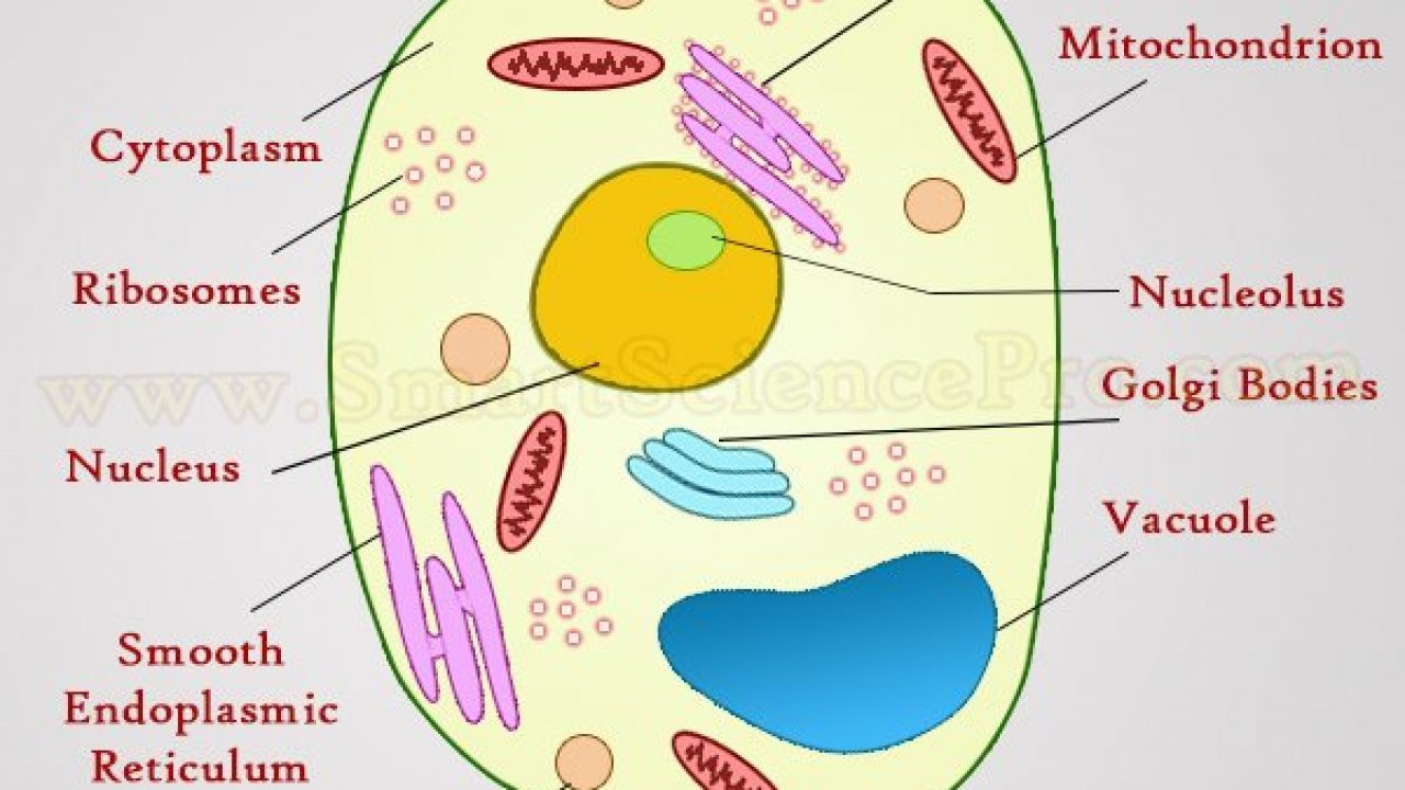
A generalised animal cell as observed under an electron microscope. 4) cell membrane or plasma membrane. Under the microscope, an animal cell shows many different parts called organelles, that work together to keep the cell functional. I am studying a bachelor's degree in biology, and i take a lot of pictures of what we do in the labs so i can remember what was everything like when i study. Feel free to post your own, but please read the rules first (see below), and note that we are not a (1a) no screenshots or pics where the only focus is a screen. They reveal complex patterns in creatures such as mice and fruit flies that are also. To look at a cell close up we need a microscope. Wide collections of all kinds of labels pictures online. Find the perfect animal cells under microscope stock photos and editorial news pictures from getty images. Animal cells under microscope stock photo c vlad nikon 110611304. Meiosis animal cell under microscope education stock photo edit. The cells may be removed directly or by mechanical or enzymatic action. As being single layers, such cells can be transferred directly to a cover slip to examine under a microscope.
Share :
Post a Comment
for "Picture Of Animal Cell Under Microscope / a. Cell : unit of function - BIOLOGY4ISC / Wide collections of all kinds of labels pictures online."


Post a Comment for "Picture Of Animal Cell Under Microscope / a. Cell : unit of function - BIOLOGY4ISC / Wide collections of all kinds of labels pictures online."